Suspicious Tumors
Suspicious tumour maybe benign or may be malignant. After biopsy it will confirm. But when anyone suspects a tumour, must consult with a doctor. Her we explain malignant tumour. Most skin Malignant Tumours are locally destructive malignant (cancerous) growth of the skin. They originate from the cells of the epidermis, the superficial layer of the skin. Unlike cutaneous malignant melanoma, the vast majority of these sorts of skin malignancy rarely spread to other parts of the body (metastasis) and become life-threatening.
Malignant lesions may cause skin changes such as acanthosis nigricans and dermatomyositis or produce secondary deposits. Lymphomas can arise in or invade the skin. Pruritus may be associated with Hodgkin’s disease.
There are three major types of skin malignancy:
- Basal cell carcinoma (the most common),
- Squamous cell carcinoma (the second most common), which originate from skin cells, and
- Melanoma, which originates from the pigment-producing skin cells (melanocytes) but is less common, though more dangerous, than the first two varieties.
- Other rare forms of skin malignancy include lymphomas, Merkel cell carcinoma, and carcinoma of other tissue in the skin, including sarcomas as well as hair and sweat gland tumours.
The following types of Suspicious tumors are tracked by Tibot:
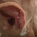
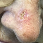
BASAL CELL CARCINOMA
KERATOACANTHOMA
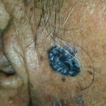
MELANOMA
SOLAR KERATOSES
SQUAMOUS CELL CARCINOMA
Squamous cell carcinoma may be painful. Both forms of skin malignancy may appear as a sore that bleeds, oozes, crusts, or otherwise will not heal. They begin as a slowly growing bump on the skin. Skin lesion may bleed after minor trauma. Both kinds of skin malignancy may have raised edges and a central ulceration.
Symptoms of basal cell malignancy include:
- Appearance of a shiny pink, red, pearly, or translucent bump
- Pink skin growths or lesions with raised borders that are crusted in the center
- Raised reddish patch of skin that may crust or itch, but is usually not painful
- A white, yellow, or waxy area with a poorly defined border that may resemble a scar.
Symptoms of squamous cell malignant tumour include:
- Persistent, scaly red patches with irregular borders that may bleed easily
- Open sore that does not go away for weeks
- A raised growth with a rough surface that is indented in the middle
- A wart-like growth
Actinic keratoses (AK), also called solar keratoses, are:
- scaly,
- crusty lesions caused by damage from ultraviolet light, often in the facial area, scalp, and backs of the hands.
These are considered precancers because if untreated, up to 10% of actinic keratoses may develop into squamous cell carcinoma.
The reasons for malignancy are:
- increased exposure to solar ultraviolet radiation (UVR)
- an increasing elder population
- increasing exposure to an increasing number of carcinogenic substances
- an increasing number of people who are immunosuppressed.
There are several effective means of treating skin cancer. The choice of therapy depends on the location and size of the tumour, the microscopic characteristics of the cancer, and the general health of the patient.
- Topical medications: In the case of superficial basal cell carcinoma, some creams, gels, and solutions can be used, including imiquimod, which works by stimulating the body’s immune system causing it to produce interferon which attacks the cancer, and fluorouracil (5-FU), a chemotherapy drug.
- Destruction by electrodessication and curettage (EDC): The tumour area with a local anesthetic is repeatedly scraped and the edge is then cauterized. The advantage of this method is that it is fast, easy, and relatively inexpensive.
- Surgical excision: The area around the tumour with local anesthetic removed and then the wound edges are closed by suture. For very big tumours, skin grafts or flaps are needed to close the defect.
- Micrographic surgery: The site is locally anesthetized and removes the visible tumour with a small margin of normal tissue. The tissue is immediately evaluated under a microscope and areas that demonstrate residual microscopic tumour involvement are re-excised and the margins are re-examined. This cycle continues until no further tumour is seen.
- Radiation therapy: Ten to fifteen treatment sessions deliver a high dose of radiation to the tumour and a small surrounding skin area. This treatment is for those who are not a patient for surgical procedure.
- Other types of treatments for skin malignancy include:
Cryo-surgery where tissue is destroyed by freezing,
Photodynamic therapy: blue light is used to destroy the malignant tissue,
Laser surgery to vaporize the skin’s top layer and destroy lesions,
Oral medication: Vismodegib, Sonidegib.
When anyone suspects a bad tumour, must consult with a doctor as soon as possible.
First time: as soon as possible visit a Dermatologist.
Then visit an Oncologist.
After that, Doctors will decide you visit schedule.
- Oxford hand Book of medical Dermatology
- ABC Of Dermatology
- Clinical Dermatology.
Suspicious Tumors
TUI - Tibot Urgency Index

Suspicious tumour maybe benign or may be malignant. After biopsy it will confirm. But when anyone suspects a tumour, must consult with a doctor. Her we explain malignant tumour. Most skin Malignant Tumours are locally destructive malignant (cancerous) growth of the skin. They originate from the cells of the epidermis, the superficial layer of the skin. Unlike cutaneous malignant melanoma, the vast majority of these sorts of skin malignancy rarely spread to other parts of the body (metastasis) and become life-threatening.
Malignant lesions may cause skin changes such as acanthosis nigricans and dermatomyositis or produce secondary deposits. Lymphomas can arise in or invade the skin. Pruritus may be associated with Hodgkin’s disease.
There are three major types of skin malignancy:
- Basal cell carcinoma (the most common),
- Squamous cell carcinoma (the second most common), which originate from skin cells, and
- Melanoma, which originates from the pigment-producing skin cells (melanocytes) but is less common, though more dangerous, than the first two varieties.
- Other rare forms of skin malignancy include lymphomas, Merkel cell carcinoma, and carcinoma of other tissue in the skin, including sarcomas as well as hair and sweat gland tumours.
The following types of Suspicious tumors are tracked by Tibot:
BASAL CELL CARCINOMA
KERATOACANTHOMA
MELANOMA
SOLAR KERATOSES
SQUAMOUS CELL CARCINOMA
Squamous cell carcinoma may be painful. Both forms of skin malignancy may appear as a sore that bleeds, oozes, crusts, or otherwise will not heal. They begin as a slowly growing bump on the skin. Skin lesion may bleed after minor trauma. Both kinds of skin malignancy may have raised edges and a central ulceration.
Symptoms of basal cell malignancy include:
- Appearance of a shiny pink, red, pearly, or translucent bump
- Pink skin growths or lesions with raised borders that are crusted in the center
- Raised reddish patch of skin that may crust or itch, but is usually not painful
- A white, yellow, or waxy area with a poorly defined border that may resemble a scar.
Symptoms of squamous cell malignant tumour include:
- Persistent, scaly red patches with irregular borders that may bleed easily
- Open sore that does not go away for weeks
- A raised growth with a rough surface that is indented in the middle
- A wart-like growth
Actinic keratoses (AK), also called solar keratoses, are:
- scaly,
- crusty lesions caused by damage from ultraviolet light, often in the facial area, scalp, and backs of the hands.
These are considered precancers because if untreated, up to 10% of actinic keratoses may develop into squamous cell carcinoma.
The reasons for malignancy are:
- increased exposure to solar ultraviolet radiation (UVR)
- an increasing elder population
- increasing exposure to an increasing number of carcinogenic substances
- an increasing number of people who are immunosuppressed.
There are several effective means of treating skin cancer. The choice of therapy depends on the location and size of the tumour, the microscopic characteristics of the cancer, and the general health of the patient.
- Topical medications: In the case of superficial basal cell carcinoma, some creams, gels, and solutions can be used, including imiquimod, which works by stimulating the body’s immune system causing it to produce interferon which attacks the cancer, and fluorouracil (5-FU), a chemotherapy drug.
- Destruction by electrodessication and curettage (EDC): The tumour area with a local anesthetic is repeatedly scraped and the edge is then cauterized. The advantage of this method is that it is fast, easy, and relatively inexpensive.
- Surgical excision: The area around the tumour with local anesthetic removed and then the wound edges are closed by suture. For very big tumours, skin grafts or flaps are needed to close the defect.
- Micrographic surgery: The site is locally anesthetized and removes the visible tumour with a small margin of normal tissue. The tissue is immediately evaluated under a microscope and areas that demonstrate residual microscopic tumour involvement are re-excised and the margins are re-examined. This cycle continues until no further tumour is seen.
- Radiation therapy: Ten to fifteen treatment sessions deliver a high dose of radiation to the tumour and a small surrounding skin area. This treatment is for those who are not a patient for surgical procedure.
- Other types of treatments for skin malignancy include:
Cryo-surgery where tissue is destroyed by freezing,
Photodynamic therapy: blue light is used to destroy the malignant tissue,
Laser surgery to vaporize the skin’s top layer and destroy lesions,
Oral medication: Vismodegib, Sonidegib.
When anyone suspects a bad tumour, must consult with a doctor as soon as possible.
First time: as soon as possible visit a Dermatologist.
Then visit an Oncologist.
After that, Doctors will decide you visit schedule.
- Oxford hand Book of medical Dermatology
- ABC Of Dermatology
- Clinical Dermatology.
Analyze Skin Disorders
Use our AI chatbot to determine your skin conditon

Get an instant skin
analysis for free
Our analysis tool is available on the web and on mobile device!