Mastocytosis
Mastocytosis is a disorder characterized by mast cell proliferation and accumulation within various organs, most commonly the skin. This term describes the various conditions in which the skin, and occasionally other tissues, contains an excess of mast cells. All types are characterized by a tendency for the skin to wheal after being rubbed. There are two main types of mastocytosis:
- cutaneous mastocytosis, which mainly affects children – where mast cells gather in the skin, but aren’t found in large numbers elsewhere in the body
- systemic mastocytosis, which mainly affects adults – where mast cells gather in body tissues, such as the skin, internal organs and bones
The World Health Organization (WHO) classification of mastocytosis includes the following:
- Cutaneous mastocytosis – Urticaria pigmentosa, maculopapular cutaneous mastocytosis, diffuse cutaneous mastocytosis, mastocytoma of skin
- Indolent systemic mastocytosis
- Systemic mastocytosis with an associated (clonal) hematologic non–mast cell lineage disease
- Aggressive systemic mastocytosis
- Mast cell leukemia
- Mast cell sarcoma
- Extracutaneous mastocytoma
Types of cutaneous mastocytosis include solitary mastocytoma, diffuse erythrodermic mastocytosis, paucicellular mastocytosis (also termed telangiectasia macularis eruptiva perstans), and urticaria pigmentosa.
There are also several subtypes of systemic mastocytosis, depending on the symptoms.
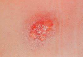
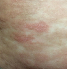
The symptoms of mastocytosis can vary depending on the type. The most common symptom of cutaneous mastocytosis is abnormal growths (lesions) on the skin, such as bumps and spots, which can form on the body and sometimes form blister.
People with mastocytosis have an increased risk of developing a severe and life-threatening allergic reaction. This is known as anaphylaxis.
The increased risk of anaphylaxis is caused by the abnormally high number of mast cells and their potential to release large amounts of histamine into the blood.
The causes of mastocytosis aren’t fully known. Mastocytosis probably is a hyperplastic response to an abnormal stimulus. Rarer cases of cutaneous mastocytosis have been associated with radiotherapy fields in patients with breast cancer.
Most cases of mastocytosis are caused by changes (mutations) in the KIT gene. This gene encodes a protein that helps control many important cellular processes such as cell growth and division, survival and movement. This protein is also important for the development of certain types of cells, including mast cells (immune cells that are important for the inflammatory response). Certain mutations in the KIT gene can leads to an overproduction of mast cells. In mastocytosis, excess mast cells accumulate in the skin and/or internal organs, leading to the many signs and symptoms of the condition.
Most cases of mastocytosis are not inherited. They occur spontaneously in families with no history of the condition and are due to somatic changes (mutations) in the KIT gene. Somatic mutations occur after conception and are only present in certain cells. Because they are not present in the germ cells (egg and sperm), they are not passed on to the next generation.
Mastocytosis can rarely affect more than one family member. In some of these cases, the condition is inherited in an autosomal dominant manner. This means that to be affected, a person only needs a change (mutation) in one copy of the responsible gene in each cell. A person with familial mastocytosis has a 50% chance with each pregnancy of passing along the altered gene to his or her child.
The KIT mutation makes the mast cells more sensitive to the effects of a signalling protein called stem cell factor. Stem cell factor plays an important role in stimulating the production and survival of certain cells, such as blood cells and mast cells, inside the bone marrow.
Conservative therapy is given. Treatment aimed at symptom relief because the prognosis for most patients with cutaneous mastocytosis is excellent. Patients advise to avoid agents that precipitate mediator release, such as aspirin, nonsteroidal anti-inflammatory drugs, codeine, morphine, alcohol, thiamine, quinine, opiates, gallamine, decamethonium, procaine, radiographic dyes, dextran, polymyxin B, scopolamine, and D-tubocurarine.
H1 and H2 antihistamines decrease pruritus, flushing, and GI symptoms. Oral disodium cromoglycate may ameliorate cutaneous symptoms, such as pruritus, whealing, and flushing, as well as systemic symptoms, such as diarrhea, abdominal pain, bone pain, and disorders of cognitive function.
The patient, premedicated with H1 and H2 antihistamines, may be started on small doses of aspirin, slowly titrated to reach a plasma level of 20-30 mg/100 mL.
Cutaneous lesions that involve a limited body area may be treated with a potent class 1 topical corticosteroid, with occlusion if required. Systemic corticosteroids are useful only in specific situations, such as ascites, malabsorption, and severe skin disease. Oral psoralen plus UV-A therapy results in general and cosmetic benefits in the treatment of cutaneous mastocytosis, particularly telangiectasia macularis eruptiva perstans. The manifestations of the disease usually recur several months after discontinuation of psoralen plus UV-A, but recurrences respond as well as the original lesions.
To confirm proper diagnosis, need to visit a consultant. Is it systemic involvement or not. If is it systemic mastocytosis then need further investigation. In systemic mastocytosis, consultation with a hematologist may be necessary for a bone marrow biopsy and staging. The WHO classification of systemic mastocytosis mandates a number of staging investigations to define the exact subtype of disease.
- Clinical Dermatology
- Roxburgh’s common skin diseases
- Andrew’s Diseases of the skin
Mastocytosis
TUI - Tibot Urgency Index

Mastocytosis is a disorder characterized by mast cell proliferation and accumulation within various organs, most commonly the skin. This term describes the various conditions in which the skin, and occasionally other tissues, contains an excess of mast cells. All types are characterized by a tendency for the skin to wheal after being rubbed. There are two main types of mastocytosis:
- cutaneous mastocytosis, which mainly affects children – where mast cells gather in the skin, but aren’t found in large numbers elsewhere in the body
- systemic mastocytosis, which mainly affects adults – where mast cells gather in body tissues, such as the skin, internal organs and bones
The World Health Organization (WHO) classification of mastocytosis includes the following:
- Cutaneous mastocytosis – Urticaria pigmentosa, maculopapular cutaneous mastocytosis, diffuse cutaneous mastocytosis, mastocytoma of skin
- Indolent systemic mastocytosis
- Systemic mastocytosis with an associated (clonal) hematologic non–mast cell lineage disease
- Aggressive systemic mastocytosis
- Mast cell leukemia
- Mast cell sarcoma
- Extracutaneous mastocytoma
Types of cutaneous mastocytosis include solitary mastocytoma, diffuse erythrodermic mastocytosis, paucicellular mastocytosis (also termed telangiectasia macularis eruptiva perstans), and urticaria pigmentosa.
There are also several subtypes of systemic mastocytosis, depending on the symptoms.
The symptoms of mastocytosis can vary depending on the type. The most common symptom of cutaneous mastocytosis is abnormal growths (lesions) on the skin, such as bumps and spots, which can form on the body and sometimes form blister.
People with mastocytosis have an increased risk of developing a severe and life-threatening allergic reaction. This is known as anaphylaxis.
The increased risk of anaphylaxis is caused by the abnormally high number of mast cells and their potential to release large amounts of histamine into the blood.
The causes of mastocytosis aren’t fully known. Mastocytosis probably is a hyperplastic response to an abnormal stimulus. Rarer cases of cutaneous mastocytosis have been associated with radiotherapy fields in patients with breast cancer.
Most cases of mastocytosis are caused by changes (mutations) in the KIT gene. This gene encodes a protein that helps control many important cellular processes such as cell growth and division, survival and movement. This protein is also important for the development of certain types of cells, including mast cells (immune cells that are important for the inflammatory response). Certain mutations in the KIT gene can leads to an overproduction of mast cells. In mastocytosis, excess mast cells accumulate in the skin and/or internal organs, leading to the many signs and symptoms of the condition.
Most cases of mastocytosis are not inherited. They occur spontaneously in families with no history of the condition and are due to somatic changes (mutations) in the KIT gene. Somatic mutations occur after conception and are only present in certain cells. Because they are not present in the germ cells (egg and sperm), they are not passed on to the next generation.
Mastocytosis can rarely affect more than one family member. In some of these cases, the condition is inherited in an autosomal dominant manner. This means that to be affected, a person only needs a change (mutation) in one copy of the responsible gene in each cell. A person with familial mastocytosis has a 50% chance with each pregnancy of passing along the altered gene to his or her child.
The KIT mutation makes the mast cells more sensitive to the effects of a signalling protein called stem cell factor. Stem cell factor plays an important role in stimulating the production and survival of certain cells, such as blood cells and mast cells, inside the bone marrow.
Conservative therapy is given. Treatment aimed at symptom relief because the prognosis for most patients with cutaneous mastocytosis is excellent. Patients advise to avoid agents that precipitate mediator release, such as aspirin, nonsteroidal anti-inflammatory drugs, codeine, morphine, alcohol, thiamine, quinine, opiates, gallamine, decamethonium, procaine, radiographic dyes, dextran, polymyxin B, scopolamine, and D-tubocurarine.
H1 and H2 antihistamines decrease pruritus, flushing, and GI symptoms. Oral disodium cromoglycate may ameliorate cutaneous symptoms, such as pruritus, whealing, and flushing, as well as systemic symptoms, such as diarrhea, abdominal pain, bone pain, and disorders of cognitive function.
The patient, premedicated with H1 and H2 antihistamines, may be started on small doses of aspirin, slowly titrated to reach a plasma level of 20-30 mg/100 mL.
Cutaneous lesions that involve a limited body area may be treated with a potent class 1 topical corticosteroid, with occlusion if required. Systemic corticosteroids are useful only in specific situations, such as ascites, malabsorption, and severe skin disease. Oral psoralen plus UV-A therapy results in general and cosmetic benefits in the treatment of cutaneous mastocytosis, particularly telangiectasia macularis eruptiva perstans. The manifestations of the disease usually recur several months after discontinuation of psoralen plus UV-A, but recurrences respond as well as the original lesions.
To confirm proper diagnosis, need to visit a consultant. Is it systemic involvement or not. If is it systemic mastocytosis then need further investigation. In systemic mastocytosis, consultation with a hematologist may be necessary for a bone marrow biopsy and staging. The WHO classification of systemic mastocytosis mandates a number of staging investigations to define the exact subtype of disease.
- Clinical Dermatology
- Roxburgh’s common skin diseases
- Andrew’s Diseases of the skin
Analyze Skin Disorders
Use our AI chatbot to determine your skin conditon

Get an instant skin
analysis for free
Our analysis tool is available on the web and on mobile device!