Melanocytic Naevi
Melanocytic naevi are pigmented moles. The word ‘melanocytic’ means that they are made up of the cells (melanocytes) which produce the dark pigment (melanin) that gives the skin its colour.
Melanocytic naevi are benign neoplasms or hamartomas composed of melanocytes, the pigment-producing cells that constitutively colonize the epidermis. Melanocytes are derived from the neural crest and migrate during embryogenesis to selected ectodermal sites (primarily the skin and the CNS), but also to the eyes and the ears.
Melanocytes clustered together form naevi. This type of moles varies in colour in different skin tones and they are easier to see on pink skins.
Some moles are present at birth or appear within first two years of life are known as congenital melanocytic naevi. Most develop during childhood and early adult life and are consequently called acquired melanocytic naevi.
Melanocytic naevi also known as banal naevi, tend to increase in number during the first three decades of life.
Thereafter, the number of naevi tends to decrease. New moles appearing in adulthood need to be monitored and checked if growing or changing.
Moles can be found anywhere on the skin, including on the hands and feet, genitals, eyes and scalp.
What are the symptoms of melanocytic naevi?
Usually there are no symptoms as such. Raised moles may catch on things. Moles may become sore and inflamed after trauma
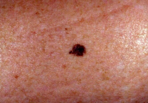
An example of what was considered a normal mole. In this case the edges were deemed to be even, not ragged and not notched. Part of the ABCDs for detection of melanoma. Source: National Cancer Institute
In general, moles are brown; however, they can come in a variety of sizes, shapes and colors:
- Shape – they can be oval and round.
- Color – they are usually medium-to dark brown, reddish brown, or flesh colored.
- Size – moles can vary enormously in size. They can cover an entire arm, or be as small as a pinhead. Typically, they are less than 6 millimeters (1/4 inch) long.
A mole’s surface can be raised, wrinkled, flat, or smooth. They may start out with one color and be flat, and then become slightly raised, and the color may lighten. Some may develop a small stalk, and gradually wear off.
Often, moles will disappear completely.
Most moles appear during early life, up to the age of about 20 years, however, they can continue appearing until middle age.
Moles that emerge after the age of 20 should be shown to a doctor. You should also see your doctor if a mole itches, has a burning sensation, is painful, bleeds or oozes, is crusty or scaly, or suddenly changes in color, elevation, size, or shape.
The cause is unknown. A genetic factor is likely in many families, working together with excessive sun exposure during childhood.
A tendency to have multiple melanocytic naevi runs in some families.
Sunburn or excessive sun exposure contribute to new moles formation and people with fair skin are more at risk.
The majority of moles are harmless and require no treatment. They may be surgically removed if:
- The mole is a suspected melanoma.
- It is bothersome, such as if the patient finds shaving difficult, or it gets snagged in clothing.
A mole may be removed in several ways:
- Shave excision – the area around the mole is numbed, a small blade is used to cut around and under the mole. A technique commonly used for smaller moles. No sutures (stitches) are needed.
- Excisional surgery (excision biopsy) – the mole plus a surrounding margin of healthy skin is cut out using a scalpel or a punch device. Sutures are required.
If the melanoma is detected in the very early stages, when the mole is thin and has not grown downwards from the surface of the skin and spread to other parts of the body, it is removed using a simple surgical technique.
If it is detected in later stages, it will be removed, plus some healthy skin around it – called a safety margin. If the cancer has entered the bloodstream or lymphatic system and formed tumors in other parts of the body, the patient will require further treatment.
- Oxford hand Book of medical Dermatology
- ABC Of Dermatology
- Clinical Dermatology
- Roxburgh’s common skin diseases
- Andrew’s Diseases of the skin
Melanocytic Naevi
TUI - Tibot Urgency Index

Melanocytic naevi are pigmented moles. The word ‘melanocytic’ means that they are made up of the cells (melanocytes) which produce the dark pigment (melanin) that gives the skin its colour.
Melanocytic naevi are benign neoplasms or hamartomas composed of melanocytes, the pigment-producing cells that constitutively colonize the epidermis. Melanocytes are derived from the neural crest and migrate during embryogenesis to selected ectodermal sites (primarily the skin and the CNS), but also to the eyes and the ears.
Melanocytes clustered together form naevi. This type of moles varies in colour in different skin tones and they are easier to see on pink skins.
Some moles are present at birth or appear within first two years of life are known as congenital melanocytic naevi. Most develop during childhood and early adult life and are consequently called acquired melanocytic naevi.
Melanocytic naevi also known as banal naevi, tend to increase in number during the first three decades of life.
Thereafter, the number of naevi tends to decrease. New moles appearing in adulthood need to be monitored and checked if growing or changing.
Moles can be found anywhere on the skin, including on the hands and feet, genitals, eyes and scalp.
What are the symptoms of melanocytic naevi?
Usually there are no symptoms as such. Raised moles may catch on things. Moles may become sore and inflamed after trauma
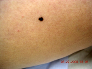
An example of what was considered a normal mole. In this case the edges were deemed to be even, not ragged and not notched. Part of the ABCDs for detection of melanoma. Source: National Cancer Institute
In general, moles are brown; however, they can come in a variety of sizes, shapes and colors:
- Shape – they can be oval and round.
- Color – they are usually medium-to dark brown, reddish brown, or flesh colored.
- Size – moles can vary enormously in size. They can cover an entire arm, or be as small as a pinhead. Typically, they are less than 6 millimeters (1/4 inch) long.
A mole’s surface can be raised, wrinkled, flat, or smooth. They may start out with one color and be flat, and then become slightly raised, and the color may lighten. Some may develop a small stalk, and gradually wear off.
Often, moles will disappear completely.
Most moles appear during early life, up to the age of about 20 years, however, they can continue appearing until middle age.
Moles that emerge after the age of 20 should be shown to a doctor. You should also see your doctor if a mole itches, has a burning sensation, is painful, bleeds or oozes, is crusty or scaly, or suddenly changes in color, elevation, size, or shape.
The cause is unknown. A genetic factor is likely in many families, working together with excessive sun exposure during childhood.
A tendency to have multiple melanocytic naevi runs in some families.
Sunburn or excessive sun exposure contribute to new moles formation and people with fair skin are more at risk.
The majority of moles are harmless and require no treatment. They may be surgically removed if:
- The mole is a suspected melanoma.
- It is bothersome, such as if the patient finds shaving difficult, or it gets snagged in clothing.
A mole may be removed in several ways:
- Shave excision – the area around the mole is numbed, a small blade is used to cut around and under the mole. A technique commonly used for smaller moles. No sutures (stitches) are needed.
- Excisional surgery (excision biopsy) – the mole plus a surrounding margin of healthy skin is cut out using a scalpel or a punch device. Sutures are required.
If the melanoma is detected in the very early stages, when the mole is thin and has not grown downwards from the surface of the skin and spread to other parts of the body, it is removed using a simple surgical technique.
If it is detected in later stages, it will be removed, plus some healthy skin around it – called a safety margin. If the cancer has entered the bloodstream or lymphatic system and formed tumors in other parts of the body, the patient will require further treatment.
- Oxford hand Book of medical Dermatology
- ABC Of Dermatology
- Clinical Dermatology
- Roxburgh’s common skin diseases
- Andrew’s Diseases of the skin
Analyze Skin Disorders
Use our AI chatbot to determine your skin conditon

Get an instant skin
analysis for free
Our analysis tool is available on the web and on mobile device!